

The indicators that drive lots of the mind and physique’s most important capabilities — consciousness, sleep, respiratory, coronary heart charge, and movement — course by bundles of “white matter” fibers within the brainstem, however imaging techniques thus far have been unable to finely resolve these essential neural cables. That has left researchers and medical doctors with little functionality to evaluate how they’re affected by trauma or neurodegeneration.
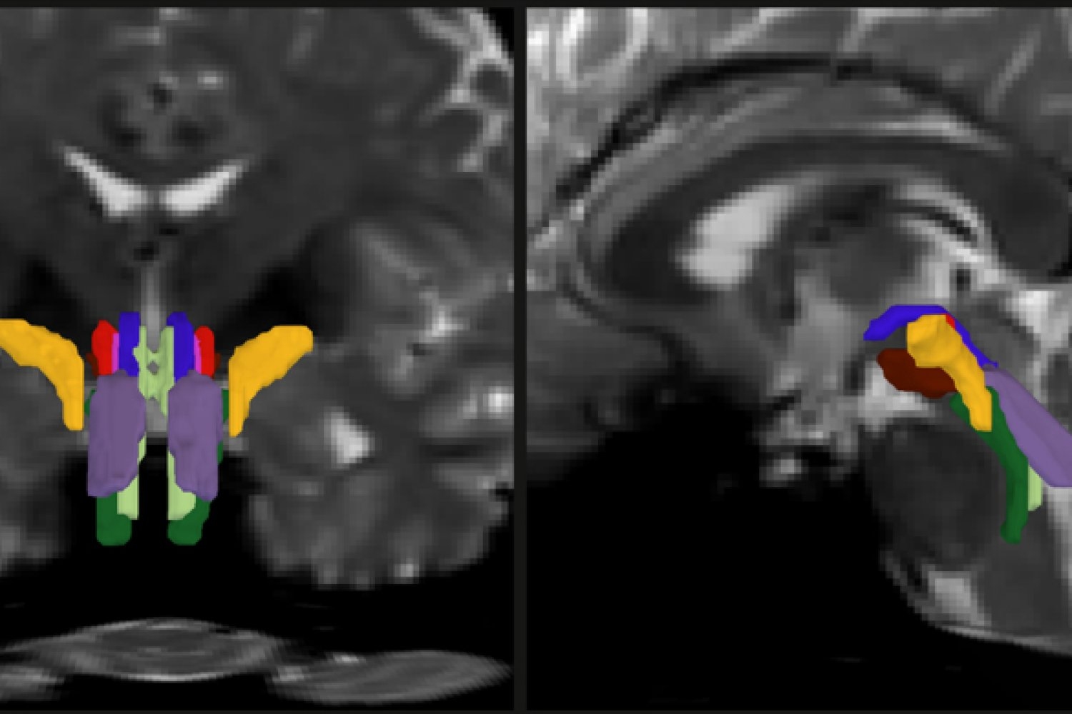
In a brand new examine, a staff of MIT, Harvard College, and Massachusetts Basic Hospital researchers unveil AI-powered software program able to mechanically segmenting eight distinct bundles in any diffusion MRI sequence.
Within the open-access examine, printed Feb. 6 within the Proceedings of the Nationwide Academy Sciences, the analysis staff led by MIT graduate scholar Mark Olchanyi stories that their BrainStem Bundle Instrument (BSBT), which they’ve made publicly obtainable, revealed distinct patterns of structural adjustments in sufferers with Parkinson’s illness, a number of sclerosis, and traumatic mind damage, and make clear Alzheimer’s illness as effectively. Furthermore, the examine reveals, BSBT retrospectively enabled monitoring of bundle therapeutic in a coma affected person that mirrored the affected person’s seven-month street to restoration.
“The brainstem is a area of the mind that’s primarily not explored as a result of it’s powerful to picture,” says Olchanyi, a doctoral candidate in MIT’s Medical Engineering and Medical Physics Program. “Individuals do not actually perceive its make-up from an imaging perspective. We have to perceive what the group of the white matter is in people and the way this group breaks down in sure problems.”
Provides Professor Emery N. Brown, Olchanyi’s thesis supervisor and co-senior creator of the examine, “the brainstem is without doubt one of the physique’s most necessary management facilities. Mark’s algorithms are a big contribution to imaging analysis and to our potential to the perceive regulation of basic physiology. By enhancing our capability to picture the brainstem, he provides us new entry to very important physiological capabilities reminiscent of management of the respiratory and cardiovascular techniques, temperature regulation, how we keep awake throughout the day and the way sleep at night time.”
Brown is the Edward Hood Taplin Professor of Computational Neuroscience and Medical Engineering in The Picower Institute for Studying and Reminiscence, the Institute for Medical Engineering and Science, and the Division of Mind and Cognitive Sciences at MIT. He’s additionally an anesthesiologist at MGH and a professor at Harvard Medical College.
Constructing the algorithm
Diffusion MRI helps hint the lengthy branches, or “axons,” that neurons lengthen to speak with one another. Axons are usually clad in a sheath of fats known as myelin, and water diffuses alongside the axons inside the myelin, which can also be known as the mind’s “white matter.” Diffusion MRI can spotlight this very directed displacement of water. However segmenting the distinct bundles of axons within the brainstem has proved difficult, as a result of they’re small and masked by flows of mind fluids and the motions produced by respiratory and coronary heart beats.
As a part of his thesis work to raised perceive the neural mechanisms that underpin consciousness, Olchanyi needed to develop an AI algorithm to beat these obstacles. BSBT works by tracing fiber bundles that plunge into the brainstem from neighboring areas greater within the mind, such because the thalamus and the cerebellum, to provide a “probabilistic fiber map.” A synthetic intelligence module known as a “convolutional neural community” then combines the map with a number of channels of imaging info from inside the brainstem to tell apart eight particular person bundles.
To coach the neural community to phase the bundles, Olchanyi “confirmed” it 30 reside diffusion MRI scans from volunteers within the Human Connectome Venture (HCP). The scans had been manually annotated to show the neural community the best way to determine the bundles. Then he validated BSBT by testing its output in opposition to “floor fact” dissections of autopsy human brains the place the bundles had been effectively delineated through microscopic inspection or very gradual however ultra-high-resolution imaging. After coaching, BSBT grew to become proficient in mechanically figuring out the eight distinct fiber bundles in new scans.
In an experiment to check its consistency and reliability, Olchanyi tasked BSBT with discovering the bundles in 40 volunteers who underwent separate scans two months aside. In every case, the instrument was capable of finding the identical bundles in the identical sufferers in every of their two scans. Olchanyi additionally examined BSBT with a number of datasets (not simply the HCP), and even inspected how every element of the neural community contributed to BSBT’s evaluation by hobbling them one after the other.
“We put the neural community by the wringer,” Olchanyi says. “We needed to make it possible for it’s truly doing these believable segmentations and it’s leveraging every of its particular person parts in a means that improves the accuracy.”
Potential novel biomarkers
As soon as the algorithm was correctly skilled and validated, the analysis staff moved on to testing whether or not the flexibility to phase distinct fiber bundles in diffusion MRI scans may allow monitoring of how every bundle’s quantity and construction diversified with illness or damage, making a novel sort of biomarker. Though the brainstem has been troublesome to look at intimately, many research present that neurodegenerative ailments have an effect on the brainstem, typically early on of their development.
Olchanyi, Brown and their co-authors utilized BSBT to scores of datasets of diffusion MRI scans from sufferers with Alzheimer’s, Parkinson’s, MS, and traumatic mind damage (TBI). Sufferers had been in comparison with controls and typically to themselves over time. Within the scans, the instrument measured bundle quantity and “fractional anisotropy,” (FA) which tracks how a lot water is flowing alongside the myelinated axons versus how a lot is diffusing in different instructions, a proxy for white matter structural integrity.
In every situation, the instrument discovered constant patterns of adjustments within the bundles. Whereas just one bundle confirmed important decline in Alzheimer’s, in Parkinson’s the instrument revealed a discount in FA in three of the eight bundles. It additionally revealed quantity loss in one other bundle in sufferers between a baseline scan and a two-year follow-up. Sufferers with MS confirmed their biggest FA reductions in 4 bundles and quantity loss in three. In the meantime, TBI sufferers didn’t present important quantity loss in any bundles, however FA reductions had been obvious within the majority of bundles.
Testing within the examine confirmed that BSBT proved extra correct than different classifier strategies in discriminating between sufferers with well being circumstances versus controls.
BSBT, subsequently, may be “a key adjunct that aids present diagnostic imaging strategies by offering a fine-grained evaluation of brainstem white matter construction and, in some instances, longitudinal info,” the authors wrote.
Lastly, within the case of a 29-year-old man who suffered a extreme TBI, Olchanyi utilized BSBT to a scans taken throughout the man’s seven-month coma. The instrument confirmed that the person’s brainstem bundles had been displaced, however not lower, and confirmed that over his coma, the lesions on the nerve bundles decreased by an element of three in quantity. As they healed, the bundles moved again into place as effectively.
The authors wrote that BSBT “has substantial prognostic potential by figuring out preserved brainstem bundles that may facilitate coma restoration.”
The examine’s different senior authors are Juan Eugenio Iglesias and Brian Edlow. Different co-authors are David Schreier, Jian Li, Chiara Maffei, Annabel Sorby-Adams, Hannah Kinney, Brian Healy, Holly Freeman, Jared Shless, Christophe Destrieux, and Hendry Tregidgo.
Funding for the examine got here from the Nationwide Institutes of Well being, U.S. Division of Protection, James S. McDonnell Basis, Rappaport Basis, American SidS Institute, American Mind Basis, American Academy of Neurology, Middle for Integration of Drugs and Revolutionary Expertise, Blueprint for Neuroscience Analysis, and Massachusetts Life Sciences Middle.