

Preparation and characterization of HMCN/Au@Hb
Initially, HMCN was fabricated as a core–shell construction utilizing a modified hard-templating methodology. Scanning electron microscopy (SEM) pictures revealed that HMCN have been spherical, uniformly distributed, and had a median diameter of 57.06 ± 10.2 nm (Fig. 1a). Transmission electron microscopy (TEM) additional demonstrated that the HMCN possessed an interior hole construction alongside with a median ultrathin shell thickness of two.35 nm (Fig. 1b). Brunauer Emmet Teller (BET) N2 absorption-desorption evaluation confirmed the presence of considerable voids with pore dimension of 1.76 nm and a excessive particular floor space as much as 532.7475 m2/g, contributing to the improved loading capability of HMCN (Fig. 1c and Determine S1). After bodily absorption of Au onto the floor of HMCN, Au offered as protrusions in TEM pictures, like chocolate beans on cookies. Au and HMCN have been thus tightly built-in and HMCN/Au was efficiently fabricated (Fig. 1d). Moreover, the lattice airplane spacing of 0.235 nm measured by means of the high-resolution TEM (HRTEM) picture corresponds to the (111) diffraction airplane of Au (Fig. 1d insert). Concurrently, the profitable ornament of Au was proved by mapping aspect profiles (Fig. 1e), and the coexistence of Au and C parts within the HMCN/Au have been additional validated by energy-dispersive X-ray spectrometer (EDS) (Fig. 1f). Afterwards, X-ray photoelectron spectroscopy (XPS) was carried out to measure the aspect composition and valence states of HMCN/Au (Fig. 1g). The high-resolution Au 4f spectrum could be deconvoluted into two doublets centered at 87.82 and 84.14 eV, labeled as Au4f5/2 and Au4f7/2, respectively, which indicated that Au precursor was partially lowered to metallic Au (Fig. 1h) [36]. As well as, the chosen space electron diffraction (SAED) sample with the (111), (200), (220) and (311) planes of cubic HMCN/Au echo the outcomes from HRTEM (Determine S2).
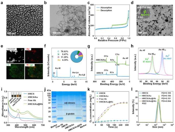
Preparation and characterization of HMCN/Au@Hb. (a) SEM and (b) TEM pictures of HMCN. (c) N2 absorption/desorption isotherms of HMCN. (d) TEM pictures of HMCN/Au (Insert was HRTEM). (e) HAADF-STEM picture and elemental mapping for HMCN/Au. (f) Quantitative elemental evaluation of HMCN/Au carried out utilizing EDS. The inserts have been the comprised aspect and corresponding atomic frequencies. (g) XPS evaluation of the HMCN and HMCN/Au. (h) The high-resolution XPS spectra of Au 4f for HMCN/Au. (i) UV-VIS spectra of free Hb, HMCN, HMCN/Au and HMCN/Au@Hb, respectively (Insert was the colour change earlier than and after loading). (j) Coomassie blue staining carried out on electrophoresed acrylamide loaded with Hb. (ok) Oxygen releasing properties measured by RDPP probe. (l) Dimension distribution of HMCN, HMCN/Au, HMCN/Au@Hb and PEGylated HMCN/Au@Hb decided by DLS.
Given to the hole and mesopores of HMCN/Au, Hb was effectively loaded and HMCN/Au@Hb was ultimately shaped. The UV-VIS spectra of HMCN/Au@Hb exhibited attribute absorption peaks at 410 and 525 nm equivalent to Hb and Au (Fig. 1i). Furthermore, the insert picture illustrates the colour adjustments through the synthesis of HMCN/Au@Hb, the darkish grey HMCNs became a barely brownish darkish gray coloration after loaded with Hb (Fig. 1i insert). Coomassie blue staining revealed protein bands at 14 and 25 kDa, in correlation with the β-globin and α/β dimers of Hb (Fig. 1j) [37]. Based mostly on the usual focus curve of Hb (Determine S3a, b), the optimum mass ratio was decided to be 1:5 (Hb: HMCN) (Desk S1). The oxygen-carrying capability of various nanomaterials was then assessed utilizing a dissolved-oxygen meter. Specifically, a major rise in O2 ranges was detected in free Hb and HMCN/Au@Hb, which confirmed that Hb retained its oxygen-carrying exercise through the synthesis course of (Fig. 1ok). Furthermore, the floor of HMCN/Au@Hb have been modified with DSPE-PEG2000-NH2 to boost the biocompatibility and stability in physiological situation. Dynamic gentle scattering (DLS) evaluation confirmed a barely elevated particle dimension following Au ornament, Hb loading and PEGylation. The hydrodynamic sizes of HMCN, HMCN/Au, HMCN/Au@Hb and PEGylated HMCN/Au@Hb dispersed in deionized water have been measured as 177.57 ± 5.10 nm (PDI = 0.105), 205.50 ± 2.50 nm (PDI = 0.106), 228.67 ± 3.05 nm (PDI = 0.151) and 259.77 ± 4.95 nm (PDI = 0.064), respectively (Fig. 1l), with a uniform dimension distribution. In abstract, the profitable preparation of HMCN/Au@Hb was confirmed by means of aspect composition dedication, morphological remark and spectral characterization.
Microwave responsive and oxygen carrying capacities of HMCN/Au@Hb
Contemplating HMCN displays excessive dielectric constants attributable to its distinctive porous construction and conductive carbon framework, the microwave-thermal efficiency of HMCN/Au@Hb was monitored underneath managed microwave parameters, together with energy density and nanocomposites focus [38]. Following a 5-min microwave irradiation (3.0 W), the management and free Hb group solely elevated to 55.5 and 57.2℃, respectively. Whereas underneath the identical irradiation situation, HMCN, HMCN/Au and HMCN/Au@Hb (400 µg/mL) have been heated as much as 60.2, 62.6 and 63.7℃, respectively, the ultimate temperature of HMCN/Au@Hb was about 8.2℃ larger than the temperature of PBS. These outcomes show that HMCN/Au@Hb contributed to enhancing microwave-to-heat conversion effectivity to a sure extent (Fig. 2a) (corresponding infrared thermal picture was offered in Fig. 2b). Moreover, HMCN/Au@Hb was irradiated with microwave underneath completely different energy densities, the temperature and the microwave energy density have been positively correlated (Fig. 2c) (corresponding infrared thermal picture was offered in Determine S4a). Furthermore, upon publicity to microwave (3.0 W), the temperature elevated from 51.3 to 53.4, 56.1, 58.4 and 62.0℃ with growing focus of HMCN/Au@Hb (0, 200, 400, 600 and 800 µg/mL) (Fig. 2d), indicating that the microwave-thermal impact of HMCN/Au@Hb was proportional to the pattern focus (corresponding infrared thermal picture was offered in Determine S4b). These findings spotlight the potential of HMCN/Au@Hb as a wonderful microwave absorption agent to enhance the microwave-thermal conversion effectivity for tumors.

Microwave responsive and oxygen carrying capacities of HMCN/Au@Hb. (a) Actual-time infrared thermal pictures and (b) corresponding heating profiles of various samples (400 µg/mL) underneath microwave irradiation (3 W, 5 min). (c) Microwave heating profiles of PBS and HMCN/Au@Hb (400 µg/mL) underneath microwave irradiation at completely different energy densities for five min. (d) Microwave heating profiles of HMCN/Au@Hb at completely different concentrations underneath microwave irradiation (3 W, 5 min). (e) ROS detection of various samples (200 µg/mL) no matter microwave irradiation (7 W, 5 min). (f) ROS detection of various samples (200 µg/mL) underneath microwave irradiation (5 min) at completely different energy ranges. (g) ROS detection of HMCN/Au@Hb suspensions at completely different concentrations underneath microwave irradiation (3 W, 5 min). (h) ROS detection of various samples (PBS, HMCN/Au and HMCN/Au@Hb) on the focus of 200 µg/mL underneath hypoxic or normoxic situations underneath microwave irradiation (3 W, 5 min). (i) Fluorescence pictures and (j) quantitative analyses of various samples exhibiting oxygen technology underneath completely different situations. (ok) Dynamic oxygen technology of various samples underneath microwave irradiation (3 W, 5 min). Information have been offered as imply values ± SD (n = 3). (* represents p<0.05, ** represents p<0.01)
Impressed by the robust SPR attribute of Au and the enough oxygen launched by Hb, we inferred that Au may generate energetic or “sizzling” electrons in order to induce electron’s transition upon microwave irradiation and additional take up oxygen molecules as sustainable gasoline for ROS technology [39]. To analyze whether or not the HMCN/Au@Hb possessed the power of microwave-dynamic efficiency, the manufacturing and species dedication of ROS underneath microwave irradiation have been carried out by means of electron spin resonance (ESR). Notably, the HMCN/Au@Hb + MW group exhibited a attribute sextet sign equivalent to superoxide anions (•O2−), which was remarkably larger than the HMCN/Au + MW group. The outcome indicated that Hb may play a significant function in oxygen-amplified microwave-dynamic efficiency (Determine S5). We then used the DCFH-DA probe to detect complete ROS technology of HMCN/Au@Hb. Upon microwave irradiation (7.0 W), the fluorescence depth of HMCN/Au@Hb was nearly 1.7 occasions larger than that of HMCN/Au, indicating the constructive impact of oxygen in amplifying ROS technology (Fig. 2e). Subsequently, the fluorescence depth of PBS, HMCN/Au and HMCN/Au@Hb confirmed a equally power-dependent improve. Below the identical energy density, the HMCN/Au@Hb group exhibited larger fluorescence depth in comparison with that of the HMCN/Au group (Fig. 2f). Furthermore, the ROS technology of HMCN/Au@Hb confirmed a concentration-dependent pattern (Fig. 2g). Subsequently, we believed that the oxygen carried by HMCN/Au@Hb facilitated the amplification of ROS technology. After irradiated with 3.0 W microwave, the ROS alerts of HMCN/Au in normoxic setting was 1.7 occasions larger than that in hypoxic setting, however akin to HMCN/Au@Hb in the identical hypoxic setting (Fig. 2h). The outcome demonstrated that Hb may complement the O2 content material in hypoxic setting, which might additional facilitate the oxidation course of.
To additional discover the interior oxygen-carrying property of HMCN/Au@Hb, we employed an oxygen indicator, RDPP probe, which could be quenched by molecular oxygen, to visually examine the oxygen manufacturing [40]. Notably, the fluorescence depth within the PBS and HMCN teams displayed a negligible change. Nevertheless, the fluorescence depth in each the free Hb and HMCN/Au@Hb group weakened dramatically, suggesting the equally superior O2 technology actions of each the free Hb and HMCN/Au@Hb (Fig. 2i), which was in keeping with the corresponding quantitative knowledge offered in Fig. 2j. In addition to, each PBS and HMCN/Au contained little or no oxygen and remained unchanged after irradiating with microwave, whereas HMCN/Au@Hb (3 mg/mL) confirmed quick improve within the O2 focus at a hypoxic answer and maintained the extraordinarily excessive stage of O2 through the technique of microwave irradiation (Fig. 2ok). The above outcomes confirmed the oxygen-carrying exercise of HMCN/Au@Hb, in addition to a small quantity of oxygen consumption to generate ROS underneath the publicity of microwave.
Cytotoxicity of HMCN/Au@Hb underneath microwave irradiation
Impressed by the ROS technology property on the answer stage, we additional evaluated HMCN/Au@Hb induced ROS outbreak on the mobile stage. DCFH-DA probe was utilized to visualise and characterize the ROS technology by way of confocal laser scanning microscope (CLSM) [41]. Herein, invisible inexperienced fluorescence was detected in cells handled with PBS or NPs alone, whereas extra intense inexperienced fluorescence was proven upon microwave irradiation. Furthermore, the brightest inexperienced fluorescence was noticed within the HMCN/Au@Hb + MW group (Fig. 3a). Moreover, the quantitative knowledge throughout completely different teams discovered that the ROS ranges within the HMCN/Au@Hb + MW group have been roughly 1.8-fold larger than that of the HMCN/Au + MW group, indicating that intrinsic oxygen-carrying capability of Hb may induce a tremendously enhanced microwave-dynamic impact (Determine S6a). Subsequently, the aforementioned cells have been harvested for movement cytometry evaluation, and confirmed a constant pattern in comparison with these noticed in DCFH-DA assay (Determine S6b). These findings straight highlighted the effectiveness of HMCN/Au@Hb in producing ROS triggered by microwave irradiation. We additional utilized higher transwell chamber to imitate “transition zone” to research whether or not tumor cells within the marginal “transition zone” may nonetheless trigger ROS burst underneath ultra-low microwave irradiation, Though the temperature within the higher chamber of the microwave irradiation group was considerably decrease than that within the backside chamber and direct heating group, the ROS ranges have been akin to these within the direct heating group. It additional demonstrated that even underneath low-power microwave irradiation within the “transition zone”, nanoparticles may nonetheless be successfully excited to provide ROS (Determine S7).

In vitro microwave-induced cytotoxic impact. (a) CLSM pictures of intracellular ROS stage with completely different remedies (inexperienced, fluorescence of DCFH-DA). (b) Cytotoxicity of Huh-7 cells handled with completely different samples and (c) handled with HMCN/Au@Hb at completely different concentrations. (d) Western blot demonstrating the HIF-1α expression ranges with completely different remedies. (e) CLSM pictures of intracellular O2 stage with completely different remedies (blue, fluorescence of Hoechst 33342; pink, fluorescence of RDPP; overlay pictures). (f) Schematic illustration of HMCN/Au@Hb mediated microwave thermal-dynamic impact. (g) Dwell/Lifeless co-staining and (h) Annexin V/PI staining of Huh-7 cells after completely different remedies. Information have been offered as imply values ± SD (n = 3). (* represents p<0.05, ** represents p<0.01)
On condition that extreme oxidative stress detrimentally impacts the mobile killing impact, the antitumor impact of HMCN/Au@Hb in Huh-7 cells was initially characterised by MTT assay [42]. After being irradiated with microwave, the cell viability fee reveals an roughly 80% discount in MW + HMCN/Au@Hb (50 µg/mL) group, which was larger than that in MW + HMCN/Au (50 µg/mL) group (Fig. 3b). Particularly, cell viability was step by step lowered within the HMCN/Au@Hb + MW group with growing concentrations of HMCN/Au@Hb, supporting its successfully antitumor impact in a concentration-dependent method. In contrast, insignificant cytotoxic impact was noticed in non-irradiated situations, suggesting the noncytotoxic nature of HMCN/Au@Hb (Fig. 3c). To additional examine the cytotoxicity of HMCN/Au@Hb in regular cell traces, human regular hepatic cell (LO2 and MIHA) and renal cell (293T) have been used within the following experiments. Even at a particularly excessive focus of 400 µg/mL, 3 forms of cell line remained viability of 76.4%, 85.6% and 80.7%, respectively, indicating the commendable biosafety of HMCN/Au@Hb (Determine S8).
Because of the enough O2 launched by HMCN/Au@Hb, we speculated that the inherent oxygen ranges would considerably elevate and mitigate the constraints imposed by the hypoxic microenvironment, thereby facilitating ROS manufacturing and amplifying the therapeutic impact [43]. Preliminary investigation of the hypoxia-relieving impact in vitro, western blot was carried out to detect the expression stage of HIF-1α [44]. HIF-1α expression stage was considerably down-regulated within the HMCN/Au@Hb group however akin to that of the unfavorable management (PBS underneath normoxic situations), solidly verifying that each Hb and HMCN/Au@Hb possessed equal hypoxia-allaying results (Fig. 3d). Subsequently, to research the dynamic technique of oxygen variation, mobile O2 content material was analyzed by CLSM remark utilizing the RDPP indicator. After incubated with HMCN/Au@Hb, the pink fluorescence depth was weakened acutely in comparison with that of the PBS group, suggesting a considerable improve in mobile oxygen ranges. Nonetheless, the pink fluorescence within the HMCN/Au@Hb + MW group was barely stronger than that within the HMCN/Au@Hb group (Fig. 3e). Fluorescence semi-quantitative (Determine S9a) and movement cytometry measurements (Determine S9b) additional confirmed that oxygen consumption might happen by means of producing ROS upon microwave stimulus. Altogether, owing to the particular hypoxic nature of tumor tissue, we speculated that the enough oxygen launched by Hb-loaded HMCN/Au may facilitate ROS technology in vitro, thereby ameliorating the hypoxic microenvironment and augmenting the therapeutic efficacy (Fig. 3f) [45].
As well as, the antitumor impact induced by extreme ROS and microwave hyperthermia was assessed utilizing Calcein-AM/PI co-staining. Ultralow cell demise charges have been noticed in non-irradiated teams. After irradiation, cell demise charges elevated dramatically, and cells incubated with HMCN/Au@Hb exhibited the very best demise fee, which was conformed to the findings of the MTT assay (Fig. 3g). The fluorescence quantitative outcomes additionally remarkably emphasised the numerous outcomes of the MW coupling system (Determine. S10). Consequently, cell apoptosis assay was carried out on Huh-7 cells to determine the kind of apoptosis triggered by ROS and hyperthermia. Cells handled with HMCN/Au + MW and HMCN/Au@Hb + MW displayed a considerable improve within the proportion of apoptotic (Annexin V-positive) and necroptotic cells (PI-positive) (Fig. 3h). Particularly, apparent apoptosis alerts (Q2 + This fall quadrant: 39.1%) have been noticed within the HMCN/Au@Hb + MW group, which was superior to that of the HMCN/Au + MW group (Q2 + This fall quadrant: 26.7%) (Determine. S11).
In vivo antitumor impact
Because of the outstanding in vitro cell demise impact, the in vivo antitumor results have been evaluated on tumor-bearing mice. The HCC fashions have been randomly divided into six teams (n = 6): PBS, HMCN/Au, HMCN/Au@Hb and their respective MW teams. Determine 4a confirmed the whole in vivo experimental course of. All through the therapy, in vivo infrared thermal pictures (Fig. 4b) and corresponding temperature curves (Fig. 4c) revealed that tumor temperature within the MW + HMCN/Au and MW + HMCN/Au@Hb teams elevated quickly inside 5 min, even reached 58.1 °C and 57.6 °C underneath an ultralow microwave energy density (3.0 W), respectively. Nevertheless, the temperature of tumors with out NPs administration solely elevated to 47.7 °C, indicating that HMCN/Au and HMCN/Au@Hb exhibited a wonderful microwave-thermal impact in vivo attributable to their related localized warmth accumulation skill. To additional consider the biodistribution of HMCN/Au@Hb, ICG labeled fluorescence imaging was carried out on Hepa1-6 tumor xenograft mice. Following intravenous injection of free ICG or HMCN/Au@Hb@ICG, fluorescence pictures have been acquired at completely different timepoints (0, 1, 2, 4, 6, 8, 12 and 24 h). Each teams exhibited robust fluorescence sign at 1 h (Determine S12a). Notably, HMCN/Au@Hb demonstrated considerably larger fluorescence retention on the tumor web site in comparison with free ICG, with secure tumor fluorescence persisting past 24 h, whereas the ICG group confirmed minimal sign past 8 h. This extended tumor accumulation was probably attributed to the optimum nanoparticle dimension, facilitating the improved permeability and retention (EPR) impact (Determine S12b). Ex vivo imaging additional confirmed sustained fluorescence in tumors, supporting the extended retention of HMCN/Au@Hb in tumor tissue (Determine S12c). As well as, we supplemented the Au content material in tumor and different organs with Inductively Coupled Plasma Mass Spectrometry (ICP-MS), it revealed that apparent Au enrichment within the tumor of HMCN/Au@Hb group on the time of sacrifice (1-hour post-injection), demonstrating the wonderful tumor accumulation potential. In addition to, it additionally demonstrated vital hepatic and renal uptake, presumably because of the major involvement of the liver and kidneys within the metabolic clearance of the nanomaterials (Determine S13). Nevertheless, ex vivo distribution of each free ICG and ICG-labeled HMCN/Au@Hb decreased quickly and was almost fully cleared after 24 h administration, as beforehand proven in Determine S12c.

In vivo antitumor impact. (a) Schematic illustration of the experimental schedule for the subcutaneous tumor mannequin remedies. (b) Infrared thermal pictures and (c) the corresponding heating profiles of tumor-bearing mice throughout microwave irradiation. Histological evaluation of sacrificed tumor tissues after a 21-day therapy (d) ROS, (e) TUNEL, (f) Ki-67 and (g) H&E staining. (h) Particular person of tumor development curves in several teams. (i) Pictures (j) and the corresponding weight of tumor tissues at 21st day after remedies. Information have been offered as imply values ± SD (n = 6). (* represents p<0.05, ** represents p<0.01)
The potent antitumor functionality of HMCN/Au@Hb was additional investigated by means of ROS, TUNEL, Ki-67 and H&E staining of tumor tissues dissociated on day 21. Important variations within the morphology of tumors have been noticed throughout completely different therapy teams. Remarkably, HMCN/Au@Hb + MW handled tumors exhibited larger ranges of ROS technology than the opposite teams, indicating that ROS outbreak might play a pivotal function in tumor suppression (Fig. 4d, Determine S14a). As well as, TUNEL staining confirmed that HMCN/Au@Hb + MW group exhibited essentially the most intense pink fluorescence, indicative of apoptotic tumor cell, highlighting the superior microwave-dynamic property to induce most cancers cell apoptosis (Fig. 4e, Determine S14b). Equally, tumors stained with Ki-67 (Fig. 4f, Determine S14c) and H&E (Fig. 4g) revealed that HMCN/Au@Hb + MW displayed the bottom tumor cell proliferation and essentially the most in depth karyorrhectic particles, in sharp distinction to the considerable proliferation and restricted necrosis noticed in different teams. These findings demonstrated that HMCN/Au@Hb achieved pronounced antitumor impact by inducing ROS outbreak upon microwave irradiation.
Inspired by the good efficiency of HMCN/Au@Hb in thermal imaging and tumor cell injury in vivo, adjustments in tumor quantity have been monitored all through 21-day therapy (Determine S15). In comparison with the quickly tumor development within the management, HMCN/Au, HMCN/Au@Hb and MW-only teams, solely reasonable tumor inhibition noticed within the HMCN/Au + MW group, whereas the tumor development curves started to extend beginning on the fifteenth day, because the inadequate oxygen might produce insufficient microwave-dynamic impact and in the end result in incomplete tumor necrosis and recurrence. In the meantime, the HMCN/Au@Hb + MW group offered with substantial tumor suppression, in addition to a major and definitive healing impact (Fig. 4h and Determine S16). Tumor pictures (Fig. 4i) and tumor weights (Fig. 4j) defined the same end in a extra intuitive and clear means. Moreover, the potential uncomfortable side effects have been evaluated through the 21-day remark interval. Notably, no vital fluctuations have been noticed in physique weight throughout all various teams, which preliminarily confirmed the wonderful biosafety of all remedies in mice (Determine S17). To additional examine potential thermal injury to regular tissues adjoining to tumor lesions post-ablation, we carried out pathological examinations. Histological analyses revealed a well-demarcated ablation zone, and there have been no apparent heating injury results in surrounding muscle tissues upon microwave irradiation (Determine S18). Furthermore, H&E staining was carried out on main organs throughout completely different handled mice, and no observable tissue necrosis was discovered. It steered that each nanomaterials administration and microwave irradiation had no seen pathological abnormalities or inflammatory lesions in vivo (Determine S19). Collectively, HMCN/Au@Hb exhibited superior tumoricidal impact and favorable biosafety, which was anticipated to be a biocompatible nanomaterial for microwave ablative remedy sooner or later.
To analyze the tumor recurrence, we additional carried out a varied of experiment teams to constantly monitor the tumor development for 33 days (Observe: tumor quantity ≥ 1500 mm3 outlined as useless). Comparative evaluation at day 7 post-ablation revealed that the MW + HMCN/Au@Hb group (V/V0 = 0.998) confirmed higher tumor inhibition impact than the MW-alone group (V/V0 = 1.555). Notably, some chosen tumor-bearing mice even achieved full tumor ablation in MW + HMCN/Au@Hb group. In distinction, the tumor quantity within the non-ablation group surpassed the moral endpoint threshold (≥ 1500 mm3), necessitating termination of remark attributable to fast development (Determine S20a). Extra encouragingly, there was no vital native recurrence within the MW + HMCN/Au@Hb group even at day 33 post-ablation (V/V0 = 0.414), in sharp distinction to the MW group (V/V0 = 7.481) (Determine S20b). As well as, the survival fee of mice handled with HMCN/Au@Hb plus MW was considerably extended (Determine S20c). Subsequently, in comparison with microwave ablation alone, HMCN/Au@Hb considerably enhanced the efficacy of microwave ablation and successfully inhibited tumor recurrence at post-ablation.
Antitumor immune response
To additional examine the underlying mechanism of the antitumor impact of HMCN/Au@Hb, the activation of systemic immunity was explored [46]. Earlier research have proven that DCs, largely contained in tumor draining lymph nodes (TDLNs), acted as potent antigen-presenting cells (APCs) for capturing tumor related antigens (TAAs) [47]. Herein, we discovered that the HMCN/Au@Hb + MW group confirmed a larger matured DCs (CD80+CD86+) infiltration, which far exceeded that noticed within the HMCN/Au + MW group (Fig. 5a). Quantitative knowledge additional confirmed these findings (Determine S21a). The outcomes implied that Hb-loaded HMCN/Au mediated microwave-dynamic impact appeared to facilitate antigen presentation to DCs and set off downstream immune response. Given to its necessary function in selling DC maturation, the differentiation of T lymphocytes inside the major tumor have been examined [48]. Notably, the larger CD8+ T cells infiltration have been evoked by microwave stimulus and HMCN/Au@Hb administration (Fig. 5b), and the upper intratumoral CD8+/CD3+ T cell ratios have been noticed in irradiated teams in contrast with non-irradiated teams, significantly together with HMCN/Au@Hb (Determine S21b). Subsequent, we carried out movement cytometry evaluation in splenic homogenates of mice to research the activation of T cells in immune organs. The share of CD8+ T cells have been considerably elevated within the HMCN/Au@Hb + MW group in comparison with the opposite teams (Fig. 5c). Likewise, larger CD8+/CD3+ T cell ratio have been noticed within the HMCN/Au@Hb + MW group than that of the HMCN/Au + MW group (Determine S21c). These findings steered that the oxygen-amplified microwave-dynamic impact enabled infiltration of immune cells to a bigger extent.

Antitumor immune response. (a) Circulation cytometry of DC maturation in TDLNs with indicated remedies. (b) Circulation cytometry of T-cell activation in tumors and (c) spleens with indicated remedies. (d) ELISA evaluation of the TNF-α, IFN-γ, IL-6, and IL-12p70 ranges in serum and tumors. Immunofluorescence pictures of (e) HIF-1α, (f) Pimonidazole and CD31+, (g) CD8, IFN-γ and Granzyme B publicity in tumors. Information have been all offered as imply values ± SD (n = 3). (* represents p<0.05, ** represents p<0.01)
Furthermore, the function of varied cell-secreted pro-inflammatory components, corresponding to TNF-𝛼, IFN-𝛾, IL-6, and IL-12p70, all of which had a significant operate in modulating immune responses, have been additional explored by way of ELISA assay [49]. ELISA assay portrayed an elevated serum secretion ranges of 1.28-, 1.68-, 1.61- and 1.33-fold for TNF-𝛼, IFN-𝛾, IL-6 and IL-12p70 in HMCN/Au@Hb + MW group than that within the HMCN/Au + MW group, respectively. As for intratumoral secretion ranges, HMCN/Au@Hb upon microwave irradiation yielded the best TNF-𝛼, IFN-𝛾, IL-6 and IL-12p70 secretions, whereas their respective secretions in different teams have been comparatively decrease, confirming that HMCN/Au@Hb plus MW may exert a strong activated-inflammatory impact (Fig. 5d).
Aiming to deepen our comprehension of the interplay between hypoxic reduction and intrinsic antitumor immunity of HMCN/Au@Hb in vivo, immunofluorescence experiments have been then carried out on mouse tumor tissues. The particular hypoxic standing of tumors was evaluated by HIF-1α expression ranges [50]. Apparently, a major down-regulation was noticed within the HMCN/Au@Hb + MW group in comparison with the HMCN/Au + MW group (Fig. 5e, Determine S22). In the meantime, accumulating proof has proven that inadequate microwave therapy may worsen hypoxia and induce angiogenesis in tumor websites, pimodazole and CD31+ staining have been adopted for detecting hypoxic areas and microvessels [51]. In contrast with the MW group, a dramatically discount of inexperienced fluorescence (pimodazole staining) and pink fluorescence (CD31+ staining) have been noticed within the HMCN/Au@Hb + MW group (Fig. 5f, Determine S22). It steered the improved oxygenation and fewer angiogenesis on the tumor web site, which might doubtlessly contribute to reversing the immune-suppressive TME. Immunofluorescence evaluation for CD8+, IFN-γ and Granzyme B additional verified the elevated infiltration of effector cytotoxic CD8+ T lymphocytes in tumor websites [47]. CD8+, IFN-γ and Granzyme B infiltration together remedy teams have been larger than that of the monotherapy teams, and the HMCN/Au@Hb + MW group confirmed the strongest fluorescence intense (Fig. 5g, Determine S22). These findings confirmed the effectivity of HMCN/Au@Hb in changing naïve T cells into effector T cells (CD8+, IFN-γ and Granzyme B). Total, the above findings demonstrated that intrinsic oxygen-carrying capability of Hb induced a tremendously enhanced microwave-dynamic impact. Altogether, HMCN/Au@Hb had the distinctive skill of in situ oxygen technology and keep in vivo hypoxia-relieving impact, which advantages for reversing the immune-suppressive microenvironment. This examine may pave the best way in direction of expediting tumoricidal immunity.